
Archivo: Spike omicron mutations side
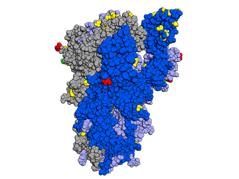
Descripción: Positions of mutations in the SARS-CoV-2 Omicron variant compared to the original strain, showing amino acid substitutions (yellow), deletions (red), and insertions (green). In this trimeric structure, two monomers (gray and light blue) have their receptor-binding domains in the "down" conformation while one (dark blue) is in the "up" or "open" conformation. "Side" view shows receptor-binding domains at the top of the image; the membrane would be located at the bottom. Mutation data source from WHO. Rendered from PDB: 6VYB. Structure, Function, and Antigenicity of the SARS-CoV-2 Spike Glycoprotein. Walls, A.C., Park, Y.J., Tortorici, M.A., Wall, A., McGuire, A.T., Veesler, D. (2020) Cell 181: 281 PubMed: 32155444 DOI: 10.1016/j.cell.2020.02.058
Título: Spike omicron mutations side
Créditos: Trabajo propio
Autor(a): Opabinia regalis
Términos de Uso: Creative Commons Attribution-Share Alike 4.0
Licencia: CC BY-SA 4.0
Enlace de Licencia: https://creativecommons.org/licenses/by-sa/4.0
¿Se exige la atribución?: Sí
Usos del archivo
La siguiente página enlaza a este archivo:
