
Archivo: Parcellation of different cortical regions involved in visual processing

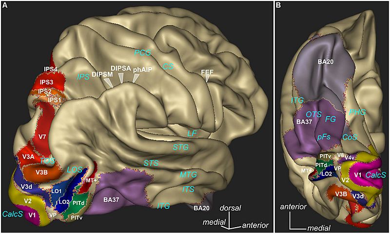
Descripción: Parcellation of different cortical regions involved in visual processing. Some of these regions are particularly involved in binocular vision and some regions are known to show deficits in amblyopes under diverse visual stimulation. Lateral view (A) and ventral view (B) are presented. The 3D rendering (Anatomist, www.brainvisa.info) represents the cortical surface of the Conte69 human surface-based atlas (Van Essen et al., 2012). V1, V2, MT+ as defined by (Fischl et al., 2008), V3A, V3B, V4v, V7, IPS1/2/3/4 as defined by (Swisher et al., 2007), V3d, LO1, LO2, PITd, PITv, as defined by (Kolster et al., 2010), occipitotemporal area BA37, inferior temporal area BA20 available in Caret software (www.nitrc.org/projects/caret/, Van Essen et al., 2001). CalcS, calcarine sulcus; LOS, lateral occipital sulcus; TOS, transverse occipital sulcus; ITG, inferior temporal gyrus; ITS, inferior temporal sulcus; MTG, middle temporal gyrus; STS, superior temporal sulcus; STG, superior temporal gyrus; LF, lateral fissure; OTS, occipitotemporal sulcus; CoS, collateral sulcus; PHG, parahippocampal gyrus; PCG, postcentral gyrus; CS, central sulcus.
Título: Parcellation of different cortical regions involved in visual processing
Créditos: Joly O and Frankó E (2014) Neuroimaging of amblyopia and binocular vision: a review. Front. Integr. Neurosci. 8:62. doi: 10.3389/fnint.2014.00062 http://journal.frontiersin.org/article/10.3389/fnint.2014.00062/full
Autor(a): Joly O and Frankó E
Términos de Uso: Creative Commons Attribution 3.0
Licencia: CC BY 3.0
Enlace de Licencia: https://creativecommons.org/licenses/by/3.0
¿Se exige la atribución?: Sí
Usos del archivo
La siguiente página enlaza a este archivo:
