
Archivo: Lung epithelium 80294-2.6
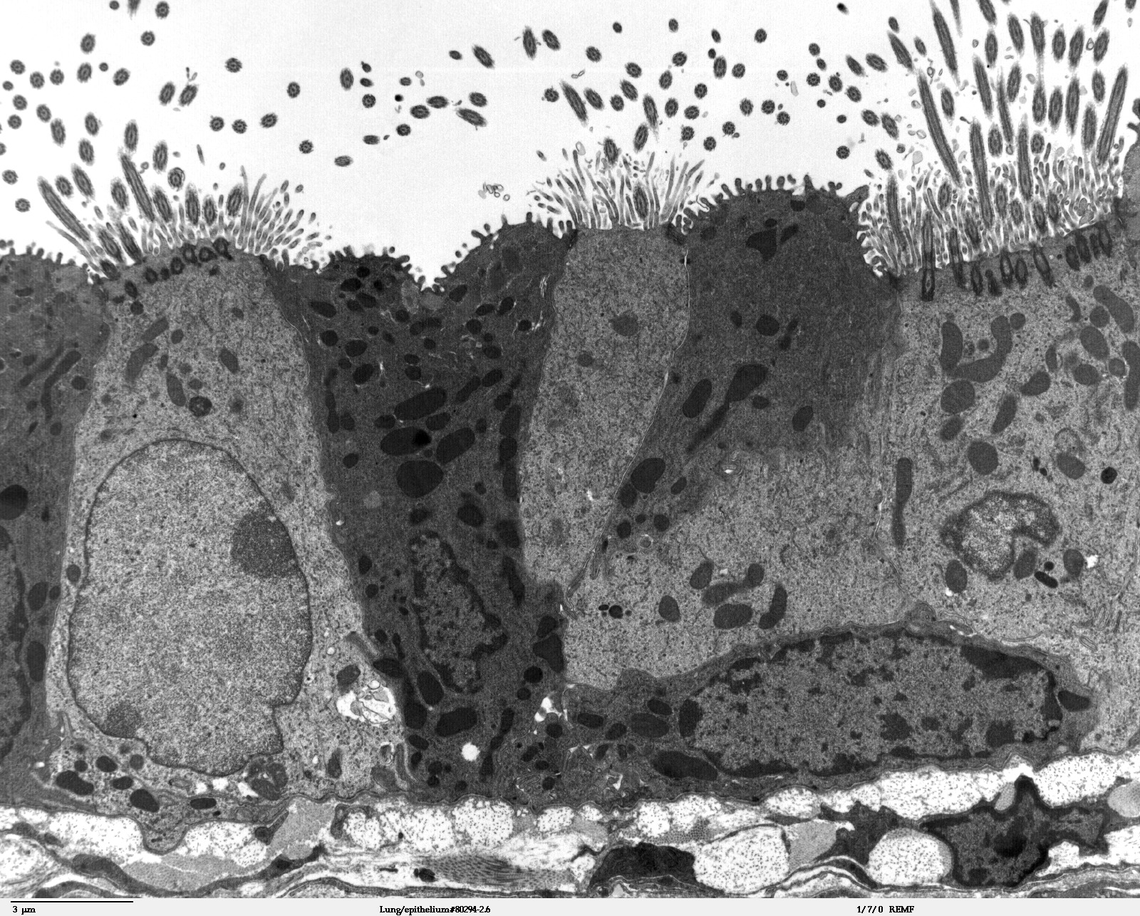
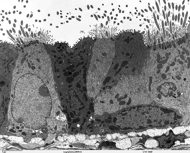
Descripción: Transmission electron microscope image of a thin section cut through the bronchiolar epithelium of the lung (mouse), which consists of ciliated cells and non-ciliated cells (called Clara cells). Image shows the ciliary microtubules in transverse and oblique section. In the cell apex are the basal bodies that are the anchoring sites for the ciliary axonemes. Note the difference in size and shape between the microvilli and the cilia. JEOL 100CX TEM
Título: Lung epithelium 80294-2.6
Créditos: http://remf.dartmouth.edu/imagesindex.html http://remf.dartmouth.edu/images/mammalianLungTEM/source/13.html
Autor(a): Louisa Howard, Michael Binder
Permiso: PD
Términos de Uso: Dominio Público
Licencia: Dominio Público
¿Se exige la atribución?: No
Usos del archivo
La siguiente página enlaza a este archivo:
