
Archivo: Image from page 230 of "The Biological bulletin"

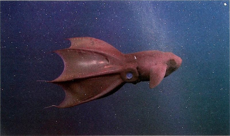
Descripción: Title: The Biological bulletin Identifier: biologicalbullet205mari Year: (s) Authors: Marine Biological Laboratory (Woods Hole, Mass. ); Marine Biological Laboratory (Woods Hole, Mass. ). Annual report 1907/08-1952; Lillie, Frank Rattray, 1870-1947; Moore, Carl Richard, 1892-; Redfield, Alfred Clarence, 1890-1983 Subjects: Biology; Zoology; Biology; Marine Biology Publisher: Woods Hole, Mass. : Marine Biological Laboratory Contributing Library: MBLWHOI Library Digitizing Sponsor: MBLWHOI Library View Book Page: Book Viewer About This Book: Catalog Entry View All Images: All Images From Book Click here to view book online to see this illustration in context in a browseable online version of this book. Text Appearing Before Image: \:\MPYROTEVTHIS BIOLUMINESCENCE 103 Text Appearing After Image: Figure 1. Vampyroteuthis infenhili.\. frame grab from high-definition video footage shot at a depth of 717 m in Monterey Bay. California. Mantle length = 10.2 cm. The fin-base photophore is located behind the dark patch of skin, posterior to (to the right of) the fin. The composite organ is the small, elongate white patch dorsal to and on a line just behind the eye. Minute epidermal organs are scattered over the surface of the mantle and the arms, but not on the web. The new arm-tip light organs described here are located on the oral surface of the filamentous portion of each arm, beyond the margin of the web. where, depending on the species (Herring, 1977). The light they produce is used for attracting prey, deterring predators, and presumably for intraspecific communication. Luminous secretions are found in a number of deep-living inverte- brates but are rare among cephalopods and fishes (Herring, 1977. 1988). Three types of light-emitting organs have been described in Vampyroteuthis infenuilis: large, paired, complex photo- phores at the bases of the fins; small, simple, epidermal organs scattered over the surface of the animal; and com- posite organs—two clusters of small, pale nodules located dorsally on a line just behind the eyes (Pickford, 1949). Light production has been observed only from the fin-base photophores; emission spectra of these organs were mea- sured at 460 nm by Herring (1983) and 461-466 nm by Widder et al. (1983). Herring et al. (1994) examined all three organ types and, based on detailed histological evi- dence, concluded that the composite organs are probably extraocular photoreceptors. while the epidermal organs are most likely light producers. They also found that the reflec- tive surfaces in the light organs were collagen instead of the iridisomal platelets found in other modern cephalopods. To date, no one has observed light from the epidermal organs, nor has there been any behavioral evidence of light sensi- tivity by the composite organs. We have discovered two new forms of bioluminescent expression in Vampyroteuthis: light produced by organs at the tips of all eight arms, and luminous fluid released by the arm tips. Materials and Methods In situ behavioral observations and quantitative video surveys of meso- and bathypelagic cephalopods have been a component of MBARI's midwater research program since 1991 (Hunt, 1996). The program is based on the use of remotely operated vehicles, or ROVs. Over a 10-year time span we have carefully observed 57 individuals of Vampy- roteuthis in situ and have collected 18 to establish in labo- ratory aquaria. Specimens in this study included adult males and females with mantle lengths ranging from 7.9 to 12.1 cm. All were gently collected with the ROV Ventana (Ro- bison, 1993) at a time-series station 1600 m deep over the axis of the Monterey Submarine Canyon. Field observations and collections occurred under full illumination from the ROVs four 500-W, broad-spectrum lights. Once the ROV was recovered, the animals were placed in darkened con- tainers and were quickly transferred to our laboratory ashore. In the shoreside facility they were maintained in the dark, at 4° to 6° C, in circular, 260-1 kreisel tanks (Hamner, 1990) for as long as 2 months. Most of the specimens appeared to be temporarily blinded by the vehicle's lights during capture. After several hours in the dark, they responded to point sources of white Note About Images Please note that these images are extracted from scanned page images that may have been digitally enhanced for readability - coloration and appearance of these illustrations may not perfectly resemble the original work.
Título: Image from page 230 of "The Biological bulletin"
Créditos: https://www.flickr.com/photos/internetarchivebookimages/20191862780/
Autor(a): Internet Archive Book Images
Términos de Uso: Creative Commons Zero, Public Domain Dedication
Licencia: CC0
Enlace de Licencia: http://creativecommons.org/publicdomain/zero/1.0/deed.en
¿Se exige la atribución?: No
Usos del archivo
La siguiente página enlaza a este archivo:
