
Archivo: A computed tomography brain scan showing bilateral basal ganglia calcification
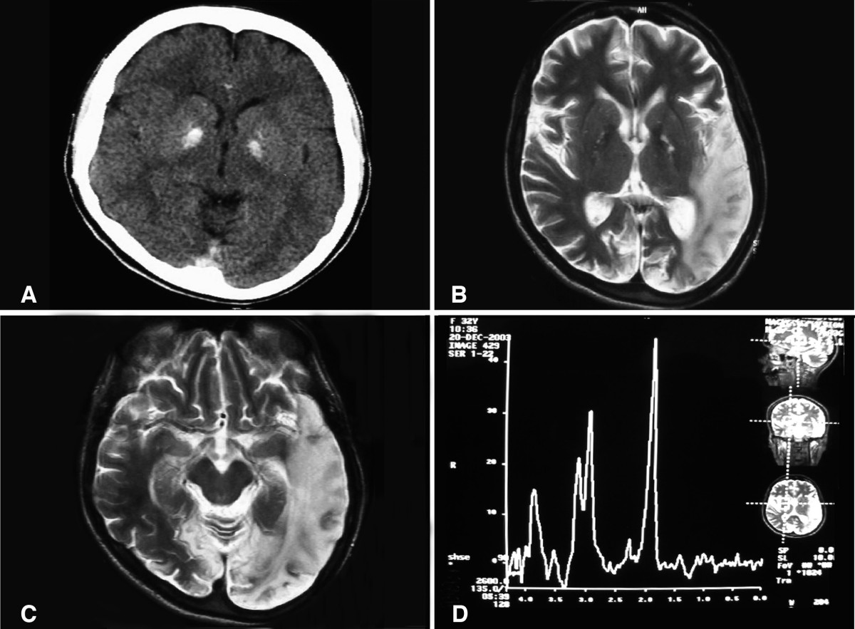
Descripción: (a) A computed tomography brain scan showing bilateral basal ganglia calcification; the cerebellum shows prominent folia indicating mild cerebellar atrophy. (b) Axial T2 brain magnetic resonance image scan showing left temporo-parieto occipital ischemic lesion. (c) Axial T2 brain magnetic resonance image scan showing the extension of the parietal temporal region to the occipital lobe, and also showing a right occipital lesion. (d) Magnetic resonance spectroscopy showing inversion of J-coupling phenomenon at 1.3 ppm, indicating lactate peak. Abu-Amero et al. Journal of Medical Case Reports 2009 3:77 doi:10.1186/1752-1947-3-77
Título: A computed tomography brain scan showing bilateral basal ganglia calcification
Créditos: A patient with typical clinical features of mitochondrial encephalopathy, lactic acidosis and stroke-like episodes (MELAS) but without an obvious genetic cause: a case report
Autor(a): Abu-Amero KK, Al-Dhalaan H, Bohlega S, Hellani A, Taylor RW.
Permiso: © 2009 Abu-Amero et al; licensee BioMed Central Ltd. This is an Open Access article distributed under the terms of the Creative Commons Attribution License (https://creativecommons.org/licenses/by/2.0), which permits unrestricted use, distribution, and reproduction in any medium, provided the original work is properly cited.
Términos de Uso: Creative Commons Attribution 2.0
Licencia: CC BY 2.0
Enlace de Licencia: https://creativecommons.org/licenses/by/2.0
¿Se exige la atribución?: Sí
Usos del archivo
La siguiente página enlaza a este archivo:
