
Archivo: Volume rendered CT scan of abdominal and pelvic blood vessels (smaller)

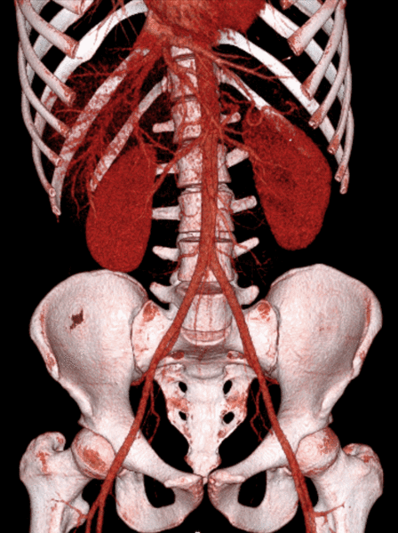
Descripción: Computed tomography of the abdomen and pelvis, performed as a contrast CT, here presented as a volume rendering of the abdominal aorta, its branches, as well as the hepatic vein. It shows normal anatomy, with no injuries. The subject is a 21 year old male who had blunt trauma to the upper abdomen during motocross.
Título: Volume rendered CT scan of abdominal and pelvic blood vessels (smaller)
Créditos: Trabajo propio
Autor(a): Mikael Häggström, M.D. Author info - Reusing images - Conflicts of interest: None Mikael Häggström, M.D. Consent note: Written informed consent was obtained from the individual, including online publication.
Términos de Uso: Creative Commons Zero, Public Domain Dedication
Licencia: CC0
Enlace de Licencia: http://creativecommons.org/publicdomain/zero/1.0/deed.en
¿Se exige la atribución?: No
Usos del archivo
La siguiente página enlaza a este archivo:
.gif&oldid=3385771)

.gif)