
Archivo: Primary biliary cirrhosis intermed mag much cropping
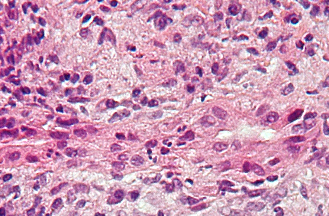
Descripción: Intermediate magnification micrograph of primary biliary cirrhosis. Liver biopsy. H&E stain. Features: Bile duct intraepithelial lymphocytes - key feature. Bile duct epithelial cells with eosinophilic cytoplasm. Granulomata - close to the bile duct. Portal inflammation with mixed cell population, i.e. lymphocytes, plasma cells, eosinophils.See also Image:primary biliary cirrhosis low mag.jpg Image:primary biliary cirrhosis intermed mag.jpg
Título: Primary biliary cirrhosis intermed mag much cropping
Créditos: Trabajo propio
Autor(a): Nephron
Términos de Uso: Creative Commons Attribution-Share Alike 3.0
Licencia: CC BY-SA 3.0
Enlace de Licencia: https://creativecommons.org/licenses/by-sa/3.0
¿Se exige la atribución?: Sí
Usos del archivo
La siguiente página enlaza a este archivo:
