
Archivo: Potential mechanisms of LTP in spinal dorsal horn in vivo
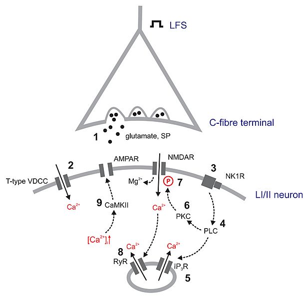
Descripción: From the article caption (CC-by 2.0): Potential mechanisms of LTP in spinal dorsal horn in vivo. At synapses between C-fibres and laminae I/II second order neurons, LFS triggers the release of the excitatory neurotransmitters glutamate and substance P leading to depolarization of the postsynaptic cell (1). T-type VDCCs are activated (2), leading to Ca2+ influx. The activation of NK1 receptors (3) activates the PLC-pathway (4), triggering the release of Ca2+ from intracellular stores (5) and the activation of PKC (6). This kinase phosphorylates NMDA receptors, thereby loosening their Mg2+ block (7). Ca2+-induced Ca2+ release activates ryanodine receptors, thereby further increasing [Ca2+]i (8). The increased [Ca2+]i is sensed by CaMKII that phosphorylates AMPA receptors, thereby potentiating synaptic strength. LFS: low frequency stimulation; SP: substance P; VDCC: voltage-dependent Ca2+ channel; AMPAR: alpha-amino-3-hydroxy-5-methyl-4-isoxazolepropionic acid receptor; NMDAR: N-methyl-D-aspartate receptor; NK1R: neurokinin receptor 1; PLC: phospholipase C; PKC: protein kinase C; IP3R: inositol 1,4,5-trisphosphate receptor; RyR: ryanodine receptor; CaMKII: Ca2+-calmodulin dependent kinase II;
Título: Potential mechanisms of LTP in spinal dorsal horn in vivo
Créditos: Drdla and Sandkühler Molecular Pain 2008 4:18 doi:10.1186/1744-8069-4-18 (http://www.molecularpain.com/content/4/1/18)
Autor(a): Ruth Drdla and Jürgen Sandkühler
Términos de Uso: Creative Commons Attribution 2.0
Licencia: CC BY 2.0
Enlace de Licencia: https://creativecommons.org/licenses/by/2.0
¿Se exige la atribución?: Sí
Usos del archivo
La siguiente página enlaza a este archivo:
