
Archivo: Polyomavirus
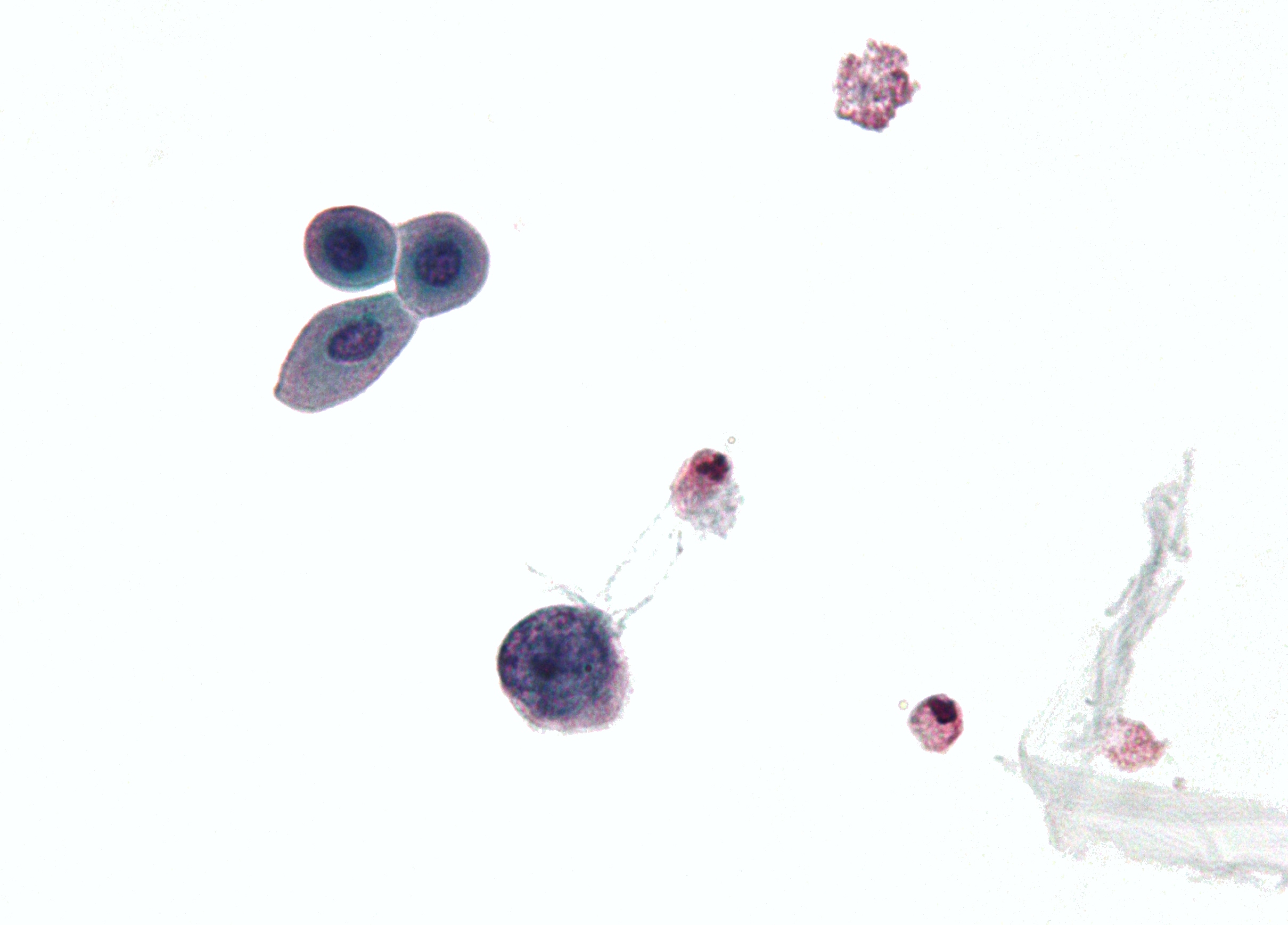
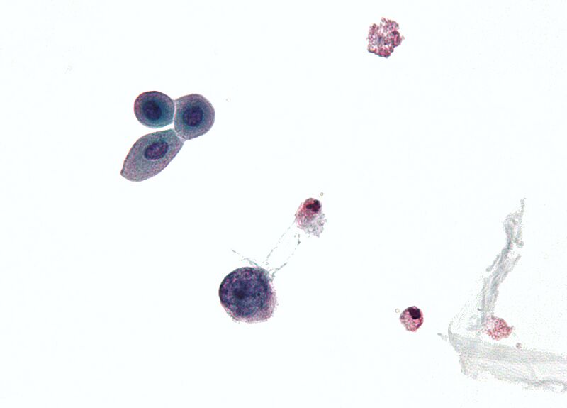
Descripción: Micrograph showing a polyomavirus infected cell. Urine cytology specimen. Features of polyomavirus infected cells (also known as Decoy cells): Usually 2X the size of a basal urothelial cell nucleus. Single cells - important feature. Scant "degenerative-appearing" cytoplasm. High NC ratio. Intranuclear inclusions - key feature. Central smudging (or "wash-out") of the chromatin/"Ground glass" chromatin. Surrounded by clear halo just deep to the nuclear membrane. Nuclear membrane clumping. See also Image:Polyomavirus 2.jpg
Título: Polyomavirus
Créditos: Trabajo propio
Autor(a): Nephron
Términos de Uso: Creative Commons Attribution-Share Alike 3.0
Licencia: CC BY-SA 3.0
Enlace de Licencia: https://creativecommons.org/licenses/by-sa/3.0
¿Se exige la atribución?: Sí
Usos del archivo
La siguiente página enlaza a este archivo:
