
Archivo: Mitosis-flourescent
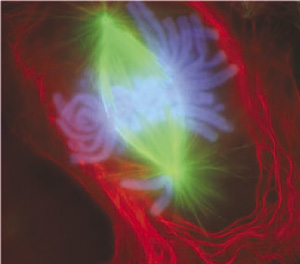
Descripción: An image of a newt lung cell stained with fluorescent dyes undergoing mitosis, specifically during early anaphase. According to NIH, "The scientists use newt lung cells in their studies because these cells are large, easy to see into, and are biochemically similar to human lung cells." The material stained green are the mitotic spindles, the material stained red is the cell membrane and some components of the cytoplasm near it, and the material stained light blue are the chromosomes.
Título: Mitosis-fluorescent
Créditos: - so it's public domain as it's US government material.
Autor(a): US government
Términos de Uso: Dominio Público
Licencia: Dominio Público
¿Se exige la atribución?: No
Usos del archivo
El siguiente archivo es un duplicado de éste (más detalles):
La siguiente página enlaza a este archivo:
