
Archivo: Histopathology of acral lentiginous melanoma in situ, intermediate magnification
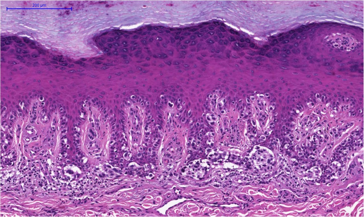
Descripción: Histopathology of acral lentiginous melanoma in situ, intermediate magnification: It shows a lymphocytic infiltrate in the papillary dermis and the irregular shape of the nests. HE stain. editar From the same case: Photography Histopathology, low magnification Histopathology, intermediate magnification Invasive
Título: Histopathology of acral lentiginous melanoma in situ, intermediate magnification
Créditos: (2015). "Focal invasiveness in complete histological analyses of a large acral lentiginous melanoma". Diagnostic Pathology 10 (1). DOI:10.1186/s13000-015-0307-z. ISSN 1746-1596. - "This is an Open Access article distributed under the terms of the Creative Commons Attribution License (https://creativecommons.org/licenses/by/4.0),"
Autor(a): Xavier-Júnior, José & Munhoz, Tania & Souza, Vinicius & Campos, Eloísa & Stolf, Hamilton & Marques, Mariângela.
Términos de Uso: Creative Commons Attribution 4.0
Licencia: CC BY 4.0
Enlace de Licencia: https://creativecommons.org/licenses/by/4.0
¿Se exige la atribución?: Sí
Usos del archivo
La siguiente página enlaza a este archivo:
